Bone Density Test Errors

In this article, my aim is to share with you two cases that came across my desk last week. Both DXA's had basic errors that could have been caught, if anyone was paying attention. Bone density is an amazing tool, and a very important piece of the puzzle when looking at bone health. This article should not make you avoid being tested. In fact, getting your first test will certainly help you and your doctors on the path to determining whether or not you have a bone density problem.
Where errors really come into play is when comparison tests are done. As you will see, in both DXA's on the next page, one of the lumbar spine and one of the femur, these types of errors can result in either reporting increased bone density or decreased bone density when a future or previous test is compared to it, unless the regional error is corrected. By the way, both errors that you see displayed below can be corrected without having a scan redone. The only time it is necessary to redo a scan on a patient is when the technician did a poor job with body placement. Frequently I send a short letter to a facility requesting changes, or give it to my patient and they will take it to the imaging facility. The changes will be made, and the imaging facility will issue a new report.
WHAT'S WRONG WITH THIS PICTURE?

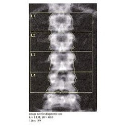
LUMBAR BONE DENSITY
The technician is responsible for setting up the patient properly and adjusting the diagnostic lines in the computer if necessary. The bone density machine sets the lines automatically to begin with, but machines can be wrong!
How the technician should be setting up the scan:
- Lumbar alignment: the technician should make sure the patient’s spine is in the middle of the scanning area, and straight.
- Analysis – line placement: The lines in the DXA machine computer demarcating L1-L4 need to be drawn properly. Knowing what is proper takes training. The bone density machine initially selects the lumbar area to be analyzed, but then the technician should make sure that the machine did the job correctly, and change or override those lines when appropriate. For example: if there is a spur that would be left out, or if there was a fracture, osteoarthritis, or surgery, these vertebra, too, would be deleted/left out of the density calculation.
- T-score L1-L4: Note that L1, 3 and 4 are within a similar range. L2 is a higher bone density. The technician should notice this, and take proper action:
If one bone in the lumbar spine is 12% lower or higher than adjacent bones, it should be removed from the calculation, and the remaining vertebra averaged for the bone density score. You need to have a minimum of two bones to be able to offer a bone density score. In the case above this did not happen – all four vertebra were included. In this case the reported bone density was off and she in fact has 2-3% lower bone density than what is reported.
Typically L1 through L4 are used when possible, and the T-score represents the bone density for the lumbar region.
Note: One standard deviation equals 12% bone density.
What the reporting doctor should have done in this case:
He/she should have deleted L2 from the diagnostic calculation, and requested images of the lumbar spine. X-rays would be the first choice. L1, L3 and L4 then should have been added together for the lumbar bone density and T-score.
What could cause one bone to have a much higher bone density than adjacent bones?
- Most commonly, osteoarthritis
- Fracture of the anterior body of L2 could also result in a higher bone density. Note that the height of the vertebrae does not look different in this one-dimensional view. However, when viewed from the side, with an x-ray, an old or new fracture could possibly present itself.
- Bone pathology could also result in a higher bone density. Certain conditions such as Paget's disease or other conditions also need to be ruled out. In the case you see here I seriously doubt that there is any bone pathology.
 Always make sure that when you get a bone density test that you get the full report, not just a two-page written report. They will often give you a CD with the images on them.
BONE DENSITY OF THE FEMUR:
The lines that you see surrounding the bones from top to bottom demarcate the analyzed area. Errors can be made if too large or too small an area is drawn, resulting in an incorrect bone density measurement.
In this particular scan, what have is the following:
Alignment: very well done.
Hip rotation: very well done.
Diagnostic area, top and bottom lines: the top line is good, but the bottom line is not placed correctly
Also noteworthy they did not include an independant T-score of the Femoral Neck
Analysis/line placement:
Note the red line at the bottom of this image. This is where the bottom line should have been placed. This error, included too much shaft of the femur that will result in a false negative of approximately 10% increase in bone density that will be included in the total score of the hip. Remember, in the hip there are two diagnostic areas: the Femur Neck, and the Total Hip. The rectangle box over the neck of the femur measures the femoral neck bone density. The total hip score includes the neck of the femur down to the bottom line that is placed on the femur.
What could happen with a false negative reading?
A follow-up DXA may show incorrect gain or loss in bone density. One possible scenario is that the next time this patient goes in to have a bone density test (typically two years later), a new technician is on board who adjusts the diagnostic area correctly. This could result in the report indicating that 10% bone loss has occurred in the hip. Anyone trained in densitometry would know that this simply does not happen over a two-year period. So, there is either an error or a potential bone pathology, such as metastatic cancer, which I have seen before. Bottom line: when there is a significant difference in bone density comparison, especially in the hip, you have to question the bone density center and ask them to take a second look; and, if you're still not satisfied, get a second opinion on your bone density from a certified bone densitometrist.
Hip Rotation:
When the hip rotation is not done properly, that can increase or decrease the bone density score by as much as 7%. As with an incorrectly drawn diagnostic box, it mostly comes into play when that scan is then compared with previous or subsequent scans, and someone is then diagnosed with either losing or gaining a significant amount of bone in the hip. A significant gain or loss should be a tip off that there is a problem with the hip rotation, or something else is wrong and further imaging is called for. It is rare, especially for people over the age of 50, to gain a significant amount of bone in the hip region.