Osteoporosis and Anorexia Athletica
February 08, 2018 6 min read

Case study: by Dr. Lani Simpson (all case studies are with permission from clients or composites).
Last year a patient came in who had recently fractured three vertebrae lifting a window. She was in a full brace from her neck to her hip to stabilize her spine. She had neverfractured a bone in her 62 years. Before her fractures she was 5’6” tall and weighed 115 pounds. The fractures were all grade 3 (about 60% loss in height of her anterior vertebral body). She said her doctors told her all lab reports were normal.
Her lab results showed low end of normal calcium of 8.6, vitamin D (29), potassium 3.4 and low end normal protein 6.3. On her history form she denied ever having had an eating disorder. Her diet appeared to be relatively healthy.
Bone density tests: (See explanation of T-scores at the end of article)
Spine L1-L4: T-score -2.5
L1 -1.5
L2 -1.0
L3 -3.9
L4 -3.6
Total Hip: T-score -3.9
Femur Neck: T-score -3.0
X-rays:
Fractures T11 (grade 2), L1 Grade 3) and L2 (Grade 2)
Two bone density tests were reviewed: Same facility, same technician and same machine. Both scans were high quality and comparable. Over the three-year period, between tests the reported loss was 5.2% bone density in the spine and 4% in the total hip region. The loss was more than the least significant change (meaning reliable).
Interview Summary:
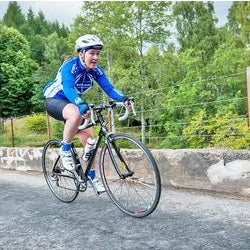
When I asked how much she ate (quantity) she noted that while she ate 3 times daily it was very small portions. I inquired about her current exercise program and she happily said she exercised daily; she rode her bike 20 miles daily prior to her fractures. She went into menopause at age 46 and did not take hormones due to a fear of breast cancer - no history of breast cancer in her family.
My assessment and working diagnosis:
Severe osteoporosis that likely began early in life. She had a form of anorexia sometimes now referred to as Anorexia Athletica. Her diet was not supporting the amount of exercise that she was doing. She noted that in her twenties her exercise was more extreme, she jogged and rode her bike for approximately 2 hours each day. She also mentioned that her period stopped for two years in her early twenties. In addition, she went into early menopause at age 46 that resulted in additional bone loss. I also suspect that her bone quality was also damaged due to a chronic lack of nutritional support for her bones.
- Recommendations: I advised her to see a top bone specialist who recommended Forteo, which I completely agreed with as her case was quite severe. Forteo not only stopped the fractures, but also healed the existing ones. I began working with her on a nutrition and supplement plan. Since this was the first time her excessive exercise was addressed, after further discussion, I recommended her to a psychotherapist who specialized in eating disorders.
- Two years later: no new fractures, no brace and she is maintaining a healthy, calorie rich diet and a balanced exercise program. Her bone density improved by 10% the first year.
Osteoporosis Definition:
Compromised bone strength predisposing to an increase risk of fracture.
Bone strength reflects the integration of two main features:
Bone density and bone quality.
Bone Density Testing Basics:
L1-L4 is the only area tested in the spine because no other bony structures are in the way, such as ribs or sacrum. When appropriate, all 4 vertebrae are added together and the T-score diagnosis is an average of all 4 vertebrae. However, when there is a significant difference between vertebrae the report should reflect that difference and make recommendations for further testing.
In this case x-rays had been taken so I knew she had fractures in the L1-L4 region. Fractured vertebrae show an increase in bone density and therefore should have been deleted. The diagnosis of bone density should have included only L3 and L4 in this case.
Note that the density of L3 and L4 is close to the total hip score. The vertebrae and total hip region contain primarily cancellous bone and that is the type of bone that women lose first in their 50s largely due to estrogen loss at menopause.
The femur neck is compact bone and loss of compact bone does not typically start to decline until around age 65 for women and 75 for men.
T-SCORE BASICS (THE FOLLOWING IS AN EXCERPT FROM, DR. LANI’S NO-NONSENSE BONE HEALTH GUIDE)
As described above, the T-score compares an individual’s bone density to the average bone density of adults of the same gender age twenty-six to twenty-nine. This comparison provides a valuable perspective for the individual being tested, as the BMD of people in their twenties is at its highest, generally speaking. The T-score gets the most attention from doctors and patients because it is the primary criterion used to diagnose low bone mass or osteoporosis, and in some cases, it serves as a justification for prescribing medications.
WHEN IS THE T-SCORE USED TO DIAGNOSE OSTEOPOROSIS?
A diagnosis of osteoporosis is made when the T-score for the lumbar spine (L-1 to L-4), neck of the femur, total hip, or forearm is –2.5 or lower in the following populations:
*women or men age fifty and over
*postmenopausal women, regardless of age
HOW ARE ETHNIC/RACIAL DIFFERENCES ACCOUNTED FOR?
When developing T-scores in the early 1990s, the World Health Organization (WHO) based its BMD measurements on those of young, healthy white women, in part because osteoporosis primarily affects Caucasian females, and there were insufficient data for the WHO to develop assessments of other ethnic groups at that time. Regardless of ethnicity, T-score calculations compare a woman’s BMD to the bone mass of Caucasian females age twenty-six to twenty-nine. Similarly, all males are compared to young Caucasian men. In general, African Americans have about 10 percent greater bone density than Caucasians, and Hispanics have about 7 percent more. Those of Asian ancestry are in roughly the same category as Caucasians in terms of bone density. However, for T-score analysis, all ethnicities are compared to either a male or female Caucasian database.
WHAT IF THE T-SCORE IS SIGNIFICANTLY LOWER THAN –2.5?
Although a T-score of –2.5 or lower is diagnostic of osteoporosis, there is a big difference between the “cutoff” score of –2.5 and scores that are much lower. Generally speaking, the farther a score falls below –2.5, the greater the cause for concern. A T-score of –4.5, for example, indicates a bone mineral density that is approximately 54 percent lower than the BMD of the control group. That is a significantly low reading; a person with a score of –4.5 is at high risk for fractures, based on bone density alone.
A diagnosis of osteoporosis is a definite red flag that should alert doctors to fully assess the patient to rule out secondary causes for the low bone density. The lower the score, the greater the possibility that a serious underlying problem (such as a nutritional deficiency or disease of the kidneys or parathyroid glands) is resulting in bone loss.
As Table 3.1 below illustrates, a T-score of –1 or better is considered normal for BMD; a reading between –1 and –2.5 indicates low bone mass, but not osteoporosis. The diagnosis of osteoporosis is assigned when the T-score is –2.5 or lower. As shown in the table’s right-hand column, each whole-unit increase or decrease in the T-score represents a 12 percent increase or decrease in BMD. Your physician may tell you that you have osteopenia. This is not a disease, but a term created by the World Health Organization (WHO) to describe low bone mass.
Table 3.1. T-Score Table
| Bone Mineral Density Level | T-score | % of BMD |
|
NORMAL BMD is no more than one standard deviation (SD) lower than the BMD of the average young adult. |
–1 or better | 12% below average or better |
|
LOW BMD (sometimes called osteopenia) BMD is between 1 and 2.5 SD below that of the average young adult. |
–1.1 to –2.4 | 13.2% to 28.8% below average |
|
OSTEOPOROSIS BMD is 2.5 or more SD below that of the average young adult. |
–2.5 and lower | 30% or more below average |
|
NORMAL (BMD is no more than one SD lower than the BMD of the average young adult.) |
–1 or better | 12% below average or better |
Note: In DXA reports or other documents related to the bone density test, while BMD readings that fall below zero have a negative or minus sign (–) in front of the number, readings above zero do not have a positive or plus sign (+) in front of them.
Occasionally, doctors, patients, and others involved in a case forget this fact and may inadvertently assume that a BMD reading without a plus or minus sign in front of it is in the negative category, creating a false diagnosis and other problems.
I once worked with a patient who was told she had low bone density because she had a T-score of 2.0. In fact, she was in the plus range. Although this is a rare occurrence, be sure to double-check any reports you receive, and ask your health care professional to verify whether your scores are on the plus or minus side.

Subscribe
Sign up to get the latest on sales, new releases and more …

