Compression Fractures of the Spine
May 20, 2025 6 min read

Compression Fractures of the Spine
Osteoporosis (weakened bone) and/or trauma contribute to compression fractures of the spine. The vast majority of compression fractures affect the spine, although they can occur in other parts of the body. Compression fractures of the spine may also be described as “vertebral fractures.” The most common cause of vertebral compression fractures is due to weakened bones. An osteoporosis fracture is one of group of conditions referred to aspathological fractures. Incidents of trauma, such as falling down stairs can result in a serious fracture. However, even with trauma the health of the bones may have been part of the fracture equation.
Compression fractures of the spine have been shown to impact how long we live. It has been reported that one significant vertebral fracture can decrease lung capacity by as much as 9%. Pneumonia is one infection that can become a problem if someone has multiple compression fractures that decrease the room for internal organs, especially the lungs. Also, pneumonia is a risk factor following a significant vertebral fracture of the thoracic spine.
Have you sustained a vertebral fracture? Did you have a silent fracture? A silent fracture is one that you did not know that you sustained. This is why if you have any previous images of your spine make sure you get the CD from the imaging facility. Those can be used to compare new imaging if there is a concern whether or not the fracture is new.
If you have ever had imaging of the spine such as x-rays, MRI’s or CT scans, the report may indicate a vertebral fracture that might also be referred to as a vertebral deformity. More importantly, the question is whether or not the fracture occurred recently. You may see the word chronic (old) or acute (new). Often it is difficult for a radiologist to determine whether or not a fracture is new on x-rays. Sometimes an MRI may be indicated to determine whether or not a fracture is recent.
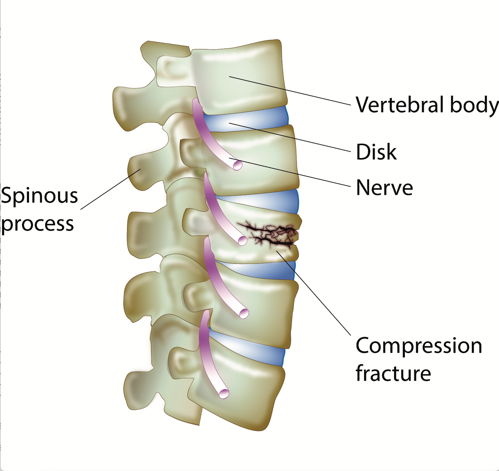
The twenty-four bones that make up the spinal column, called “vertebrae,” rest one on top of the other like a stack of boxes. With a compression fracture, typically, the front (anterior body) of the vertebra collapses to a small or large degree, possibly causing pain and loss of height in one or more vertebrae. Compression fractures of the spine occur most commonly in the thoracic (middle back) and lumbar (lower back) regions.
For patients that have been diagnosed with osteoporosis I often order baseline (standing) x-rays of the thoracic and lumbar regions. If a fracture is indeed old, often that is apparent to a radiologist due to arthritic changes surrounding the fracture.
If you want to gain more knowledge on bone health, sign up for the weekly Master Class!
We cover integrative information on bone health and have fantastic interviews with experts in the field.
What Breaks a Bone?
Most of us have seen what happens to an overloaded supermarket paper bag when the weight of the groceries puts more pressure on the bottom of the bag than it can handle. Similarly, when a bone is hit with excess force or more pressure than it can withstand, it breaks—and this is the process that is typical of most trauma fractures that result from events like a car crash or a fall off a ladder. You can see, then, why osteoporosis fractures are sometimes referred to as “minimal trauma fractures,” or “fragility fractures”—because the bones break under the minimal pressure caused by ordinary activities like stepping off a curb or even coughing. These types of fractures typically occur with advanced osteoporosis.
One statement I hear from patients goes like this, "I fractured a vertebra, but it was due to trauma." The next question is, was the health of the bone part of the reason the bone fractured? Another common trope, “I have fractured 5 bones in my life, but I am athletic." Many people are athletic without fracturing 5 bones. I am concerned about bone health when fractures are part of the story. And while bone density may be normal, remember a fracture, determined to be an osteoporosis fracture, trumps bone density alone. So, one can have normal bone density by a bone density test, yet have serious osteoporosis based on the way the bone fractured. A patient may report that they stumbled on the lawn and "shattered" their wrist. A comminuted fracture (in pieces) is of particular interest in the diagnose.
One of the methods used to measure the extent of a compression fracture is Genant’s grading system, which indicates what percentage of bone has collapsed. It classifies compression fractures as follows:

- + Grade 1 (mild)—20–25 percent bone collapse
- + Grade 2 (moderate)—25–40 percent bone collapse
- + Grade 3 (severe)—40+ percent bone collapse Small compression fractures do not always produce pain or much discomfort, and some people remain unaware that they have sustained a fracture. They are often discovered incidentally, when an X-ray of the spine is taken for another reason. Fractures seen on X-rays may have occurred in childhood, and sometimes it is hard for a radiologist to confirm if the fracture is relatively new or old. Multiple compression fractures can lead to the hunched-over posture seen in older adults, an image that is most commonly associated with osteoporosis. However, one, two, or even three mild compression fractures do not necessarily result in obvious disfigurement. An extremely hunched-over posture (Dowager’s hump) is primarily seen in very advanced cases of osteoporosis in elderly people.
On an x-ray report, you may also see a diagnosis of an end-plate fracture. That means it is less than 10% and if you fell off a horse, that fracture would indicate to me that your bones are able to withstand a solid force. It all depends on how the radiologist reports it and often, they DO NOT use the grading system. This is why I always want to view the actual x-rays myself. I have seen many errors on x-ray reporting over the years.
The most common types of spinal fractures include:
- Compression fractures: Compression fractures are small breaks or cracks in your vertebrae that are caused by traumas or develop over time as a result of osteoporosis. Osteoporosis is a disease that weakens your bones, making them more susceptible to sudden and unexpected fractures. An undiagnosed spinal compression caused by osteoporosis can make you lose several inches from your height or develop a hunched forward posture (kyphosis).
- Burst fractures:Burst fractures happen when your spine is suddenly compressed with a strong force. They can cause your vertebrae to break into many pieces.
- Chance (flexion/distraction) fractures:Chance fractures happen when your vertebrae are suddenly pulled away from each other. They’re almost like the opposite of a burst fracture.
- Insufficiency fractures: when a bone, usually the spine, sacrum or pelvis sustain a fracture simply due to body weight. Obviously, these fractures indicated severe osteoporosis with a high risk of future fractures.
When a patient sustains a compression fracture of one or more vertebrae, a doctor may offer him or her the option of undergoing some sort of surgical intervention including vertebroplasty or kyphoplasty. Each of these procedures presents its own problems and side effects; however, sometimes they are necessary, as a severe compression fracture can place pressure on a spinal nerve or on the spinal cord, resulting in serious pain. I will write more about this in the future.
In order to determine the health of bone, a bone density test fills in a big piece of the puzzle. Another piece of the puzzle is the Trabecular Bone Score (TBS) that helps determine the quality of bone. The TBS score is a test that is available at some bone density imaging centers. Lab work to diagnose bones properly is critical. Is the patient actively losing bone? One bone density test does not determine that. As you can see there is a lot of work needed to evaluate an individual set of bones. Consider learning the language of bone. To that end, my book is available at libraries or one line.
1. Lau E, Ong K, Kurtz S, Schmier J, Edidin A. Mortality following the diagnosis of a vertebral compression fracture in the Medicare population. J Bone Joint Surg Am. 2008 Jul;90(7):1479-86. doi: 10.2106/JBJS.G.00675. PMID: 18594096
2. Marek AP, Morancy JD, Chipman JG, Nygaard RM, Roach RM, Loor MM. Long-Term Functional Outcomes after Traumatic Thoracic and Lumbar Spine Fractures. Am Surg. 2018 Jan 1;84(1):20-27. PMID: 29428017.
Subscribe
Sign up to get the latest on sales, new releases and more …

