Why did this Woman Fracture her Vertebrae?
April 03, 2024 4 min read

Case Study: Vertebral Fractures
(All case studies are with permission from clients or composites)
Last year a patient who had recently fractured three vertebrae lifting a window came to me. She was in a full brace from her neck to her hip to stabilize her spine. She had never fractured a bone in her 62 years. Before her fractures she was 5’6” tall and weighed 115 pounds. The fractures were all grade 3 (about 60% loss in height of her anterior vertebral body). She said her doctors told her all lab reports were normal.
Her lab results showed low end of normal calcium of 8.3, vitamin D (29 - low), potassium 3.4 (low-end normal), Total protein 6.5 (low-end normal). Twenty-four-hour urine collection for calcium was 20 (low end normal). Her C-telopeptide (CTX) was 750 and her P1NP (procollagen type one) was 125. On her history form she denied ever having had an eating disorder. Her diet appeared to be relatively healthy, and she denied having digestive problems.
Here are her bone density test T scores:
Spine L1-L4: T-score -2.5 (borderline osteoporosis)
L1 -1.5
L2 -1.0
L3 -3.3
L4 -3.6
Total Hip: T-score -3.9
Femur Neck: T-score -3
X-rays: Fractures T11 (grade 3), L1 Grade 3) and L2 (Grade 2)
Two bone density tests were reviewed: Same facility, same technician and same machine. Both scans were high quality and comparable. Over the three-year period, between tests the reported loss was 5.2% bone density in the spine and 4% in the total hip region. The loss was more than the least significant change (meaning reliable).
Interview Notes
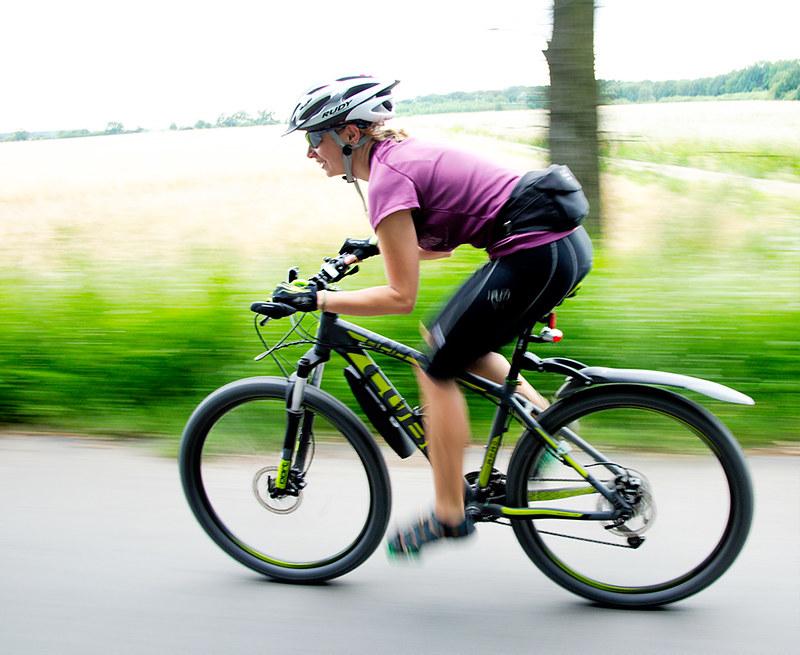
When I asked how much she ate (quantity) she noted that while she ate 3 times daily it was very small portions. I inquired about her current exercise program and she happily said she exercised daily. She rode her bike 15-20 miles daily.
She went into menopause at age 46 and did not take hormones due to a fear of breast cancer - no history of breast cancer in her family.
My assessment and working diagnosis
Severe osteoporosis that began early in life. She had a form of exercise anorexia, sometimes now referred to as Anorexia Athletica. Her diet was not supporting the amount of exercise that she was doing. She noted that in her twenties her exercise was more extreme and she jogged and road her bike for approximately 2 hours each day. She also mentioned that her period stopped for two years in her early twenties.
My recommendations
I advised her to see a top bone specialist who recommended Forteo (teriparatide), which I agreed with. Forteo not only stopped the fractures, but also healed existing fractures.
I then began working with her on a nutrition and supplement plan. Since this was the first time her excessive exercise was addressed, after further discussion, I recommended her to a psychotherapist who specialized in eating disorders.
One year later: no new fractures, no brace and she is maintaining a healthy, calorie rich diet. Her bone density improved by 10% the first year. She did have nerve pain from one fracture that resulted in nerve compression. At some point, she may need surgery for that. Otherwise, her life is back to normal.
Summary: why her bone density was so low.
- Regardless of bone density scores, her diagnosis is severe osteoporosis with high fracture risk.
- She probably did not build up a good peak bone mass by the age of 30 due to not getting enough nutrients and calories needed to build and maintain bone density
- Continued high exercise program of biking and not enough nutrients to stabilize bone
- Exercise choice: Bike riding is a great exercise however, no impact so minimal impact on bone – especially the spine. Muscle building and impact exercises are crucial.
- Estrogen loss that was not addressed at age 46.
- Lab work indicated that her nutritional status was not supporting her bone even though her lab results were within normal limits all were on the low end. Even her 24-hour urine test so 20 (should be in mid-range – around 120) along with serum calcium of 8.3 indicates low calcium intake. Protein also is needed for bone health.
- Bone turnover markers confirmed that she was actively losing bone, guesstimate about 5% yearly.
- Forteo (Teriparatide) was the right choice to stop her fractures and improve her bone density AND bone quality.
Ultimately this woman will always have a serious case of osteoporosis.
However, close monitoring her case will be the key to allowing her to have an active and physically independent life. In her case that means lifestyle changes, such as eating enough, adjusting exercise routines to include impact and weight lifting, working with a psychotherapist and bone specialist, and using the correct medications.
Over time, her needs will change, but the acceptance of her osteoporosis and willingness to change her life style choices will significantly reduce her fracture risk.
Want more information from Dr. Lani?
- Watch the 3-part video series Osteoporosis Fundamentals Bundle designed for the newly diagnosed. Currently on sale!
- Join the live weekly Masterclass for live lectures.
- Read Dr. Lani's No-Nonsense bone Health Guide.
*Note regarding bone density reports*
L1-L4 is the only area in the spine that is tested for bone density because no other bony structures are in the way of the scanner, such as ribs or sacrum. When appropriate, all 4 vertebrae are added together, and the T score is an average of all 4 vertebra. However, when there is a significant difference between one vertebra and adjacent vertebra the report should reflect that difference and make recommendations for further testing.
In this case x-rays had been taken because I suspected that she had fractures in the L1-L4 region. Fractured vertebrae show an increase in bone density and therefore must be deleted from the diagnosis. The diagnosis of bone density should have included only L3 and L4 in this case. Note that L3 and L4 density is close to the Total hip T score. The vertebra and total hip contain primarily trabecular bone and that is what women especially lose due to estrogen loss starting before menopause and 5-7 years after. The femur neck is compact bone and that does not tend to change until around age 65 for women and 75 for men.
Subscribe
Sign up to get the latest on sales, new releases and more …

